background

About Implantable Defibrillator

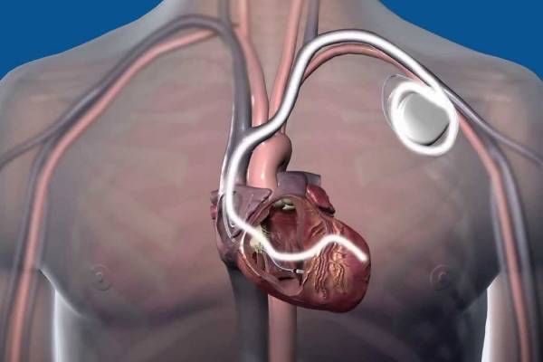
An implantable converter-defibrillator is a device that is implanted in the chest to detect possible cardiac disorders and prevent the irregular electrical activity of the heart. The defibrillator monitors the electrical conduction system of the heart and sends electric impulses to normalize the rhythm of the heart whenever needed. The implantable defibrillator responds to irregular heartbeats that arise from the lower chambers of the heart, that is, ventricles. Therefore, the device responds to Ventricular Arrhythmia, which comprises rapid and irregular heartbeats in the lower chambers of the heart. It includes premature ventricular contraction where premature heartbeats arise from the ventricles, Ventricular tachycardia where fast heart rhythms arise from the ventricles, accelerated idioventricular rhythm where the heart beats at the rate between 40 to 120 beats per minute and affects the cardiac muscle alone, Arrhythmogenic Cardiomyopathy, that primarily affects the tissues of the right ventricle, with associated irregular heartbeats. It first attacks the right ventricle, which puts a lot of stress on the left ventricle, and then, if not treated, stiffens and weakens both the ventricles. It also includes ventricular fibrillation which refers to disorganized electrical activity in the ventricles and torsades de pointes, which is a specific type of abnormal heart rhythm that leads to sudden cardiac death.

Types Of Implantable Defibrillator

There are two types of the cardiac therapy device:-

  • A traditional implantable defibrillator device - in which insulated wires or leads are implanted to the chest which attaches to the heart.
  • A subcutaneous implantable defibrillator device - which is planted under the skin at the side of the chest below the armpit. It is larger than the traditional device but doe not get implanted to the heart
Who Needs An Implantable Defibrillator
An implantable defibrillator is needed by those individuals who:-
  • Are suffering from coronary artery disease, which is a condition where the coronary arteries are narrowed and hardened due to a lack of oxygen-rich blood supply to the heart muscle by the cholesterol deposits in the walls of the coronary arteries. This results in the death of the heart muscle, leading to a heart attack or gradually progressing to heart failure. This device is also needed by those individuals who suffer from the acute coronary syndrome, which occurs when there is a reduction in the blood flow to the cardiac muscle that decreases its functions or leads to death of the cardiac muscle tissues, or arteriosclerosis, which is characterized by the loss of elasticity of the coronary artery walls.
  • Are suffering from ventricular fibrillation or ventricular tachycardia.
  • Are suffering from cardiomyopathy. In cardiomyopathy, blood flow to the heart muscle is restricted, which makes the heart muscle work harder, to the limit where it enlarges and stiffens, causing the death of the tissues of the muscle. Cardiomyopathy is one of the main causes of a heart attack or heart failure.
  • Have had a heart attack. This happens when blood supply to the heart suddenly stops, which happens due to plaque (cholesterol) deposits in the walls of the coronary artery, which block the passage of blood flow to the heart muscle. If the blood supply to a heart muscle is very low, it ruptures the entire pumping system of the heart, which includes blood vessels, arteries, capillaries, and veins.
  • Have suffered from cardiac arrest, which happens when there is a sudden loss of blood in the body due to failure of the heart to pump blood. It requires immediate action like performing CPR to restore the lack of oxygen in the body.
  • Are suffering from Brugada syndrome, which is caused when the electrical signals that help the individual cells of the heart to communicate, are disrupted, leading to a genetic mutation in the person's DNA. Symptoms include blacking out and sudden cardiac death.
  • Are suffering from Long QT syndrome, which is a heartbeat disorder causing abnormal heart rhythms.
  • Are suffering from congenital heart disease. This can lead to heart defects in infants, which might progress to a possible heart failure in the future. If caused by genes, it results in disorders like Turner's syndrome, Holt-Oram syndrome, Di George syndrome, Noonan syndrome, Alagille syndrome. If caused due to environmental factors, causes disorders in the infant are caused due to unhealthy practices carried out by the mother like alcohol consumption, smoking, antidepressants, and so on.
What To Expect Before The Procedure
Before you are ready for implantation, your doctor might need reports of tests like:-
  • Electrocardiography - electrocardiography measures the electrical activity of the heart by placing electrodes on the skin. This detects the minute changes that take place as a result of the contraction and relaxation of the cardiac muscle. This helps to check the electrical activity of the heart and arrhythmia.
  • Echocardiogram - In this procedure, a device called a transducer is placed on the top of the chest and ultrasonic waves are sent to the heart bounce off and show the live image of the heart, using a monitor.
  • Stress Test - in many cases, athletes face a heart attack or even sudden death because their hearts cannot withstand the strenuous and vigorous activities they engage in. To avoid this situation, the cardiac stress test is done where their heart activity is monitored while they are undergoing vigorous training or while running on a treadmill. Cardiac stress tests like these are done by exercise physiologists under the supervision of a sports cardiologist.
  • Remote Patient Monitoring - with the evolution of technology, advanced devices have contributed to improving the quality of the patient's life. Devices like blood pressure cuff, glucometer consists of sensors. If the device detects any abnormality in the blood pressure levels, the data is immediately transmitted to the healthcare providers. In this way, the healthcare providers are immediately aware of the patient's condition and take necessary steps accordingly.
  • Electrophysiology study - This test is done to check abnormal heart rhythms. The patient is supposed to do this test with an empty stomach, which also includes that the patient cannot take any medicines that they have been prescribed before the test begins. The electrophysiology study (EPS) is done by inserting a sheath at the artery or vein. Through the sheath, the specialized catheters with be directed towards the heart. Electric impulses will be sent through the catheters which the heart will receive and send its electric signals. The catheters pick these signals which are recorded and seen with the help of the video screen attached to it. This procedure is also called cardiac mapping and it is done with athletes to check whether they have abnormal heart rhythms which is one of the main causes of myocardial infarction

After the doctor gets the reports, you have to follow some measures before the procedure:-

  • The physician will ask you to not take certain medicines like aspirin, anti-inflammatory drugs, or blood thinners before the procedure. You need to mention the medicines that you have been given by your doctor, to the physician.
  • You should not eat or drink six to eight hours before the procedure.
  • Gather all the medicines that have been approved by the physician
  • You will be asked to remove any jewelry from your body. The doctor will also tell you to empty your bladder.
  • You will be placed at the back of your chest on the table. The electrical activity of your heart, your heart rate, and blood pressure will be monitored throughout the procedure.

During the procedure :

  • Large electrode pads are attached to the back and front of the chest.
  • The insertion site is cleaned with antiseptics. Then a numbing medicine is injected into the insertion area.
  • A plastic tube, called a sheath, will be inserted into the area, usually under the collarbone. Through this, the ICD wire will be inserted and advanced to the heart.
  • After the lead wire is inserted into the heart, it is checked whether it is in the appropriate place. A special type of X-ray. called Fluoroscopy is done to check the position of the lead wire.
  • The generator of the ICD will be inserted under the skin just below the collarbone through the insertion site after the lead wire is connected to the generator.
  • After the ICD generator is attached to the lead wire, its mechanism will be monitored through the electrocardiography monitor.

For a subcutaneous implantable defibrillator device:-

  • One or two incisions are made near the upper and lower position of the breast bone. The lead wire is then clipped under the skin next to the breast bone. From the breast bone, then it is taken to the incision on the left side of the chest.
  • The generator of the ICD will be inserted under the skin on the left side of the chest after the lead wire is connected to the generator.
  • The device is inserted on the left side of the chest near the heart.
What To Expect After The Procedure
After the procedure :
  • Your blood pressure and heart rate will be checked.
  • You should try walking slowly so that you do not face any dizziness.
  • You have to wear the sling for a day or two, depending on the doctor's advice.
  • Medicines like painkillers will be given if you feel any pain or swelling at the insertion site.
  • Ask the cardiologist or the cardiac nurses about certain restrictions and precautions that you have to maintain, like not engaging in vigorous activities or not lifting any heavy object.
  • After you are discharged and return home, you can resume your normal activities
  • You have to visit your doctor every 6 months to check whether the device is working efficiently or not.
  • If you experience fever, a feeling that the generator is loose, an ICD shock, palpitations, give a call to a nearby hospital.
Risks Associated With Implantable Defibrillator
There are certain risks associated with this procedure :
  • The patient might have allergic reactions to anesthesia
  • The patient might experience swelling, pain, or bruises in the area where the device has been placed.
  • Infection
  • While inserting the wires, the blood vessel might suffer from an injury
  • Heart rhythm problems
  • The lungs might collapse
  • The heart muscle might face an injury
  • The wires might be dislocated

The faculty of Dr. Swapnil Mate's Cardiology clinic includes experienced cardiologists, cardiac surgeons, cardiac imaging specialists, a preventive cardiology team, experienced pediatric cardiac surgeons and assisting surgeons, physical therapists, nutritionists, geneticists, child-life specialists, a multidisciplinary interventional cardiology team, and a group of pathologists who run by tests and give the proper cause of diseases. The cardiologists and surgeons recommend the ideal surgery to the patients. They mention the advantages and risks associated with the surgeries they plan to do on them. Together, they provide services that cater to their needs. Consult Dr. Swapnil Mate for the best medical assistance.

  BOOK NOW|   CALL NOW|  CHAT