background

Balloon Valvulopasty

banner

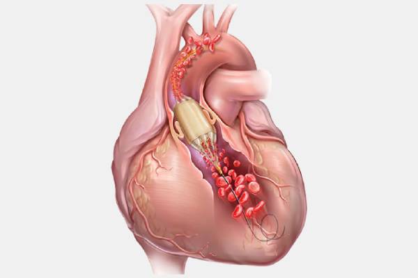
The balloon valvuloplasty is done specifically on infants who are suffering from heart disease(valvular disorders). It is done by placing a catheter through the blood vessel into the heart. The balloon is placed at the tip of the catheter. When it is blown, it forces the leaflets of the valves(cusps) to open. The balloon valvuloplasty is done in mitral, aortic, or tricuspid valves. The heart valves are responsible for sending blood to the pulmonary artery for oxygenation of the blood and transferring that oxygen-rich blood to the heart.

The Heart Valves

The heart consists of 4 valves that are responsible for the passage of blood to the heart and from the heart to other organs of the body :-

Mitral valve, also known as the bicuspid valve is one of the four valves of the heart. It is located between the upper left and lower left chambers of the heart. Also known as the bicuspid valve, the mitral valve is the only valve that has two flaps or cusps instead of three. The valve opens and closes based on the pressure differences in the upper and lower left heart chambers. The leaflet opens when there is greater pressure in the upper left chamber(left atriums) and closes when there is greater pressure on the lower left chamber (left ventricle). All of this happens within the duration of our heartbeat. The upper left chamber of the heart receives blood rich in oxygen from the pulmonary veins situated in the lungs. When the upper left heart chamber is filled with oxygen-rich blood, the mitral valve opens and blood flows to the lower left heart chamber. After transferring the blood to the lower left heart chamber, the mitral valve closes immediately to prevent regurgitation.

Also known as the right atrioventricular valve, the tricuspid valve is a boundary existing between the upper right heart chamber and the lower right heart chamber. The tricuspid valve is responsible for the transformation of deoxygenated blood into oxygen-rich blood. The blood flows from the upper right heart chamber to the lower right heart chamber and it then exits the heart through the pulmonary artery. The pulmonary artery then transfers blood to the lungs for its oxygenation. The tricuspid valve is named so because it has three flaps or leaflets or cusps that, after receiving blood from the upper right chamber of the heart (right atrium), closes immediately to prevent blood from flowing back to the right atrium, that is, it prevents tricuspid regurgitation. The tricuspid valve is located above the lower right heart chamber, on the right dorsal side of the heart.

Pulmonic valve, also known as the pulmonary valve is the valve that helps circulate blood in the body. The pulmonary valve is located between the lower right heart chamber and the pulmonary artery. The pulmonary valve opens when the pressure in the lower right heart chamber(right ventricle) rises above the pressure in the pulmonary artery and the valve closes on,y when the pressure in the right ventricle drops rapidly. Regurgitation cannot take place in the pulmonary valve since it is a unidirectional valve, that is, in the case of the pulmonary valve, blood cannot flow back into the heart.

The aortic valve is the last valve of the heart, located between the lower left chamber of the heart and the aorta. The aorta is the largest artery in the body. This is one of the most important structures of the circulatory system since it originates from the lower left heart chamber and extends to the abdomen where it transports oxygenated blood to all parts of the body. The aortic valve opens when pressure rises in the lower left heart chamber(left ventricle), allowing the passage of blood to exit from the heart to the aorta where it gets distributed to all the organs of the body. When the pressure in the left ventricle drops the aortic valve closes

Artificial Heart Valves

Living with an artificial valve in your heart does not change anything. Artificial valves work just like normal valves. They last more than 20 years. People who have gone through heart failure or either of their valves are not functioning properly, need a valve replacement. Artificial valves are mainly of three types :-

  • Mechanical valves - which come in various designs like bileaflet, caged-ball, tilting disc, and others
  • Bioprosthetic valves - that are made from animal tissues like bovine(cow) tissue and porcine(pig) tissue
  • Tissue-engineered valves - are made by cultivating human cells onto a platform. This platform acts as an extracellular matrix, directing tissue development into the right 3D design of the heart valve. Unfortunately, they are not commercially available.

In case of replacement with a mechanical valve, even though they are more durable than any other artificial valve, they can form blood clots which can cause blockage in blood vessels leading to myocardial infarction(heart attack). Patients with mechanical valves need to take anticoagulants for the rest of their life to prevent such accidents. The scenario is just the opposite concerning bioprosthetic valves. They do not form blood clots but are not durable enough. So the person with this valve has to go through another valve replacement.

Who Needs A Balloon Valvuloplasty
A balloon valvuloplasty is needed by those individuals who are suffering from :
  • Pulmonary Atresia - a condition where the pulmonary valve, the valve responsible for carrying deoxygenated blood from the heart to the lungs is not formed at all.
  • Tetralogy of Fallot - a congenital disorder comprising of four cardiac defects, pulmonary stenosis(obstruction of blood flow from the lower right heart chamber to the pulmonary artery), ventricular septal defect, right ventricular hypertrophy, overriding aorta.
  • Ebstein's Anomaly - a critical congenital heart defect in which the septal and posterior leaflets of the tricuspid valve are placed towards the apex of the right ventricle. It is commonly associated with supraventricular tachycardia.
  • Bicuspid aortic valve - is an inherited heart disease in which the third leaflet of the aortic valves fuses in the womb during development. This forms a two-leaflet aortic valve instead of the normal three-leaflet.
  • Aortic Stenosis - is a condition where the exit of the left ventricle, from where the oxygenated blood travels to the aortic valve for its transmission to the other organs, is narrowed. The narrowing may also occur at the aortic valve.
  • Mitral Stenosis - is a condition where the opening of the mitral valve is narrowed. One of the main causes of mitral stenosis is rheumatic heart disorder.
  • Aortic Regurgitation - is a condition where the aortic valve leaks that cause the blood to flow in the reverse direction, from the aorta to the left ventricle.
  • Mitral Regurgitation - is a condition where during the heart pumps out blood, the mitral valve does not close properly. This results in the backflow of blood from the left ventricle into the left atrium.
  • Tricuspid Regurgitation - is a condition where the tricuspid valve, located between the right atrium and right ventricle, does not close properly while the right ventricle contracts.
Symptoms Of Valvular Heart Disease

Symptoms include

  • Chest pain(angina) - described as a sensation of heartburn, tightness, pressure, or squeezing.
  • Shortness of breath - As the heart rate changes, the breathing pattern will also change simultaneously. If the heart is not pumping blood efficiently, this causes shortness of breath. Therefore, shortness of breath mainly arises due to low oxygen in the blood.
  • Sweating
  • Fatigues, dizziness
  • Nausea or vomiting
  • Swelling of legs, ankles, feet, abdomen due to failure of the right ventricle, which causes fluid build up in the body tissues, resulting in swelling.
  • Fatigue and weakness due to lack of oxygen in the blood
  • Irregular heartbeat or Arrhythmias due to issues in the electrical conduction system of the heart.
  • Rapid gain in weight due to fluid build-up in the body tissues
Types Of Balloon Valvuloplasty
Types of balloon valvuloplasty are:
  • Balloon aortic valvuloplasty - this procedure is used to open a narrowed aortic valve caused by aortic stenosis. It involves widening the aortic valve where calcium deposits are present. The narrowed aortic valve is widened by using a long, flexible tube (catheter) and placing it in between the deposits. The catheter is inserted into the main blood vessel of the lower arm and passed carefully into the area where an independent blood supply is given to the heart. A balloon is placed at the tip of the tube in such a way that when it is blown, it forces the valve to wide open. This allows blood to flow to the heart.
  • Balloon mitral valvuloplasty - this procedure is used to open a stiff mitral valve caused by mitral stenosis. It involves passing the catheter through the femoral vein into the right atrium(right upper heart chamber). The wall that separates the upper heart chambers is punctured and then passed to the left atrium

What To Expect Before The Procedure
Before the procedure :
  • The physician will do a thorough physical examination of you. Physical examination includes blood pressure test, triglyceride test, etc. The examiner might ask for any unusual changes that might have occurred in your body. This helps the examiner to analyze your medical history. They also analyze the patient's condition by checking the individual's height and weight. Doctors or nurse practitioners or an assistant of a physician do certain tests to analyze the condition or disease and recommend proper instructions respectively.
  • You should do certain tests before you come to the physician for your treatment. Some tests include electrocardiography which detects the minute changes that take place as a result of the contraction and relaxation of the cardiac muscle, echocardiogram, where a device called a transducer is used to show the live image of the heart. Test reports of intravascular ultrasound can also be asked for, where a catheter instead of a transducer to capture the images of coronary arteries. This helps the specialists to see whether the blood flow is obstructed or not. Coronary angiography also uses a catheter but before inserting it in the body through wrists or legs, it injects a dye into the bloodstream to create X-ray images of the coronary arteries.
  • The physician will ask you to not take certain medicines like aspirin, anti-inflammatory drugs, or blood thinners before the procedure. You need to mention the medicines that you have been given by your doctor, to the physician.
  • If you have any kind of cardiovascular disease, report this to your physician.
  • If you are allergic to any medicines, substances like latex, anesthetic agents, or any kind of narcotics, inform the surgeon before the procedure.
  • If you have any kind of bleeding disorder, report this to your physician.
  • You should not eat or drink three hours before the procedure.
  • If you are pregnant or think you might be, report this to your physician.
  • Gather all the medicines that have been approved by the physician.
  • You will be asked to remove any kind of jewelry from your body.
  • You will be changed into a hospital gown. You also have to empty your bladder before the procedure
What To Expect During The Procedure
During the procedure :
  • Fluids and medicines will be given to relax your body. Anticoagulants might also be given through an intravenous catheter inserted in your arm, or hand.
  • The doctor will numb the area where the catheter has to be inserted. It can be the femoral artery(thigh), radial artery(forearm), brachial artery(upper arm). A small, thin, guidewire is inserted into the artery.
  • Throughout the procedure, your heart rate, blood pressure will be monitored through the electrocardiography monitor, ambulatory blood pressure monitor, and other monitors present in the room
  • Once the numbing medicine has done its work, an introducer will be inserted into an accessible blood vessel. This will guide the catheter to travel in the right direction.
  • The catheter will be inserted through the introducer and will be passed into the heart.
  • After the catheter is present in the correct place, a contrast dye will be flushed into the inserted site to look at the affected area. The injection of the dye will be viewed through the monitor
  • A balloon is placed at the tip of the catheter. When the balloon is inflated, you might feel some discomfort in your chest which will subside after the balloon deflates.
  • Once the valve is opened, the insertions site will be closed by using collagen to seal the exposed blood vessel
  • Your doctor may ask you to not remove the introducer for a few hours. This device helps in wearing off the effects of blood thinners.
What To Expect After The Procedure
After the procedure :
  • The nurses will take you to the recovery room where your vital signs will be monitored.
  • After 4 to 5 hours, the introducer will be removed from the insertion site.
  • Ask the cardiologist or the cardiac nurses about certain restrictions and precautions that you have to maintain, like not engaging in vigorous activities or not lifting any heavy object.
  • After you are discharged and return home, drink plenty of water to flush out the contrast dye from your body.
  • If you experience pain, swelling, change in temperature, or color of the skin, in the area where the catheter was inserted, immediately call the hospital.

The faculty of Dr. Swapnil Mate's Cardiology clinic includes experienced cardiologists, cardiac surgeons, cardiac imaging specialists, a preventive cardiology team, experienced pediatric cardiac surgeons and assisting surgeons, physical therapists, nutritionists, geneticists, child-life specialists, a multidisciplinary interventional cardiology team, and a group of pathologists who run by tests and give the proper cause of diseases. The cardiologists and surgeons recommend the ideal surgery to the patients. They mention the advantages and risks associated with the surgeries they plan to do on them. Together, they provide services that cater to their needs. Consult Dr. Swapnil Mate for the best medical assistance.

  BOOK NOW|   CALL NOW|  CHAT