
3D Echocardiography
3-D echocardiography is the method in which a three-dimensional image of the heart is obtained instead of the traditional two-dimensional image. With the use of a matrix array probe and a proper processing system, it helps capture the image of the heart chambers through different angles. This advanced procedure of echocardiography enables analyzing the heart if it is affected by congenital disorders, coronary artery diseases, or cardiomyopathies.
Types Of Echocardiography
The types of echocardiography include :-
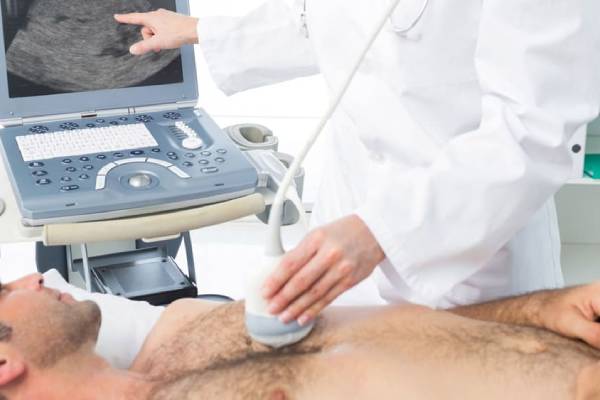
This is the standard procedure of performing echocardiography. The transducer is placed on the top of the chest and ultrasonic waves are sent to the heart bounce off and show the live image of the heart, using a monitor.
This procedure is done when a clearer image of the heart is required. The transducer is placed at the tip of a specialized probe. It is passed through the esophagus and allows the image from a location that is just behind the heart.
This procedure is taken in two phases. In the first phase, the echocardiography is done while the person is lying down. In the second phase, the procedure ( also known as the cardiac stress test) is done by monitoring the patient's heart activity while they are undergoing vigorous training or while running on a treadmill. Cardiac stress tests like these are done by exercise physiologists under the supervision of a cardiologist.
This procedure uses a catheter instead of a transducer to capture the images of coronary arteries. This helps the specialists to see whether the blood flow is obstructed or not. Coronary angiography also uses a catheter but before inserting it in the body through wrists or legs, it injects a dye into the bloodstream to create X-ray images of the coronary arteries.
This procedure uses catheters to insert the transducer inside the heart to view the 2d structures inside the area. This technique is used to cross the wall that separates the two upper chambers of the heart, to gain access to the left and right chambers of the heart.
Who Needs A 3-D Echocardiography
The types of echocardiography include :-
What To Expect Before The Procedure?
What To Expect During The Procedure?
The results of the echocardiography will be given to the cardiologist of the facility or to the patient's physician, who then, analyses the report and gives instructions to their patient accordingly.
The faculty of Dr. Swapnil Mate's Cardiology clinic includes experienced cardiologists, cardiac surgeons, cardiac imaging specialists, a preventive cardiology team, experienced pediatric cardiac surgeons and assisting surgeons, physical therapists, nutritionists, geneticists, child-life specialists, a multidisciplinary interventional cardiology team, and a group of pathologists who run by tests and give the proper cause of diseases. The cardiologists and surgeons recommend the ideal surgery to the patients. They mention the advantages and risks associated with the surgeries they plan to do on them. Together, they provide services that cater to their needs. Consult Dr. Swapnil Mate for the best medical assistance.